
Simple and affordable retinal diagnostics at home for improved and personalized AMD treatment.
Facts & Background information
Simple and affordable retinal diagnostics at home for improved and personalized AMD treatment.
Facts & Background information
AMD is the leading cause of blindness
Age-related macular degeneration (AMD) is a disease that is the leading cause of blindness in developed countries [2]. It affects the retina’s point of sharpest vision, the macula. A distinction is made between the early (dry) form of AMD and the acute (wet) form that is developed by around 15% of the patients [1]. While the dry form progresses only very slowly, it sometimes evolves into the wet form which progresses intermittently and, if left untreated, leads to a loss of central visual acuity.
How many people are affected?
Age-related macular degeneration accounts for 8.7% of all blindness worldwide and is the most common cause of blindness in developed countries [2]. In Germany, for instance, AMD is by far the most frequent cause of blindness with 49.8% [3]. While the wet AMD accounts for around 15% of all AMD cases, it is responsible for 80% of all vision loss caused by AMD [1].
Is there a treatment for AMD?
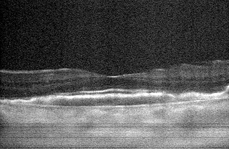
For several years, wet AMD can be effectively treated using VEGF inhibitors, which are injected into the eye. However, this treatment needs to be repeated. Optical Coherence Tomography (OCT) is used to acquire three‑dimensional images of the retina, which help to determine whether an injection is necessary.
AMD in Homecare
How are we improving AMD treatment?
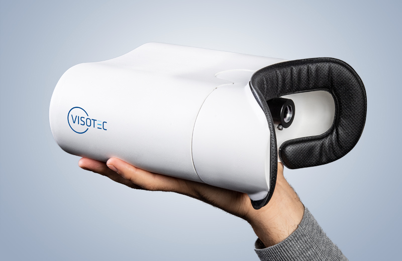
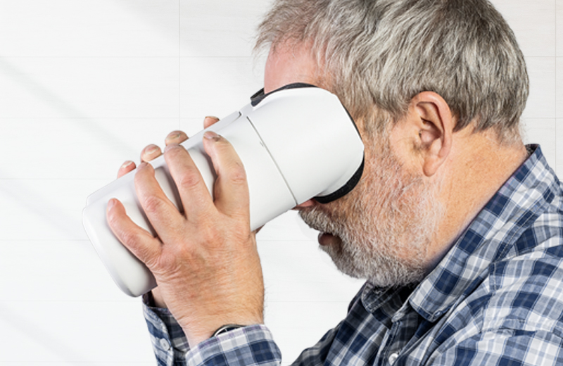
We have developed a solution that enables automated inspection of the retina with a compact, affordable, and simple-to-use device. This allows the patient to regularly check whether disease activity is present. If necessary, the patient is sent to the ophthalmologist for treatment. This prevents undertreatment and the consequent loss of vision.
What prevented this approach until now?
Current OCT systems are bulky, expensive, and are only used by trained personnel. Thus, they are mostly used in hospitals or doctors’ offices, where a regular daily or even weekly check for every patient is not feasible.
Our home care OCT technology
We have developed proprietary OCT technology that is based on a full-field approach [4, 5]. This technology enables building small and cheap handheld devices which can be used by patients to diagnose the retina by pressing a single button and looking into the device. Our technology is patented: [6, 7, 8, 9].

What others say about it
Please find statements and commens of important sources below.
Comment of the the Association for Research in Vision and Ophthalmology
(Claus von der Burchard/UKSH Kiel, Dr. Claude Burgoyne/ARVO President 2018):
Sources
[1]
Kaschke, Michael ; Donnerhacke, Karl-Heinz ; Rill, Michael S.: Optical
Devices in Ophthalmology and Optometry: Technology, Design Principles and
Clinical Applications. Wiley-VCH, 2014
[2]
Wan Ling Wong, et.al., Global prevalence of age-related macular degeneration and disease burden projection for 2020 and 2040: A systematic review and meta-analysis, The Lancet, Vol 2 February 2014, p.106–16
[3]
Finger, R.P. et.al., Incidence of blindness and severe visual impairment in Germany, Invest Opthalmol Vis Sci, 2011, 52(7); p. 4381–9
[4]
Sudkamp, Helge ; Koch, Peter ; Spahr, Hendrik ; Hillmann, Dierck ;
Franke, Gesa ; Münst, Michael ; Reinholz, Fred ; Birngruber, Reginald ; Hüttmann, Gereon: In-vivo retinal imaging with off-axis full-field
time-domain optical coherence tomography. Vol. 41, No. 21 / November 1 2016 / Optics Letters
[5]
Sudkamp, Helge ; Hillmann, Dierck ; Koch, Peter ; Vom Endt, Malte ; Spahr, Hendrik ;
Münst, Michael ; Pfäffle, Clara ; Birngruber, Reginald ; Hüttmann, Gereon: Simple approach for aberration-corrected OCT
imaging of the human retina. Vol. 43, No. 17 / 1 September 2018 / Optics Letters
[6]
Hendrik Spahr ; Helge Sudkamp ; Gereon Hüttmann ; Peter Koch ; Gesa Franke ; Dierck Hillmann ; Reginald Birngruber. Verfahren und Vorrichtung zum Ablichten wenigstens einer Schnittfläche im Innern eines Licht streuenden Objekts. DE102015113465A1.
[7]
Hillmann, Dierck ; Hüttmann, Gereon ; Koch, Peter ; Spahr, Hendrik ; Sudkamp, Helge. Vorrichtung zur ophthalmologischen Blickfixierung für Patienten mit beliebiger Sehschärfe. DE102017129951B3.
[8]
Hillmann, Dierck ; Hüttmann, Gereon ; Koch, Peter ; Spahr, Hendrik ; Sudkamp, Helge. Verfahren zum Ablichten einer Sequenz von Schnittflächen im Innern eines Licht streuenden Objekts mit verbesserter Abtastung. DE102018106292B3.
[9]
Hillmann, Dierck ; Hüttmann, Gereon ; Koch, Peter ; Michael Münst ; Spahr, Hendrik ; Sudkamp, Helge. Full-Field OCT-Verfahren und –System zum Erzeugen einer Abbildung eines Augenhintergrunds. EP000003517021A1.